Read Deep Imaging in Tissue and Biomedical Materials: Using Linear and Nonlinear Optical Methods - Lingyan Shi | ePub
Related searches:
Deep Imaging in Tissue and Biomedical Materials - Routledge
Deep Imaging in Tissue and Biomedical Materials: Using Linear and Nonlinear Optical Methods
Deep Imaging in Tissue and Biomedical Materials - Amazon.com
Deep Imaging in Tissue and Biomedical Materials Taylor & Francis
Deep Imaging in Tissue and Biomedical Materials / Shi, Lingyan
Deep Imaging in Tissue and Biomedical Materials : Using Linear
Deep Imaging in Tissue and Biomedical Materials Optics
Deep tissue fluorescence imaging and in vivo biological
Deep Imaging in Tissue and Biomedical Materials (Book) on OnBuy
Deep tissue optical focusing and optogenetic modulation with
Biomedical Imaging and Instrumentation Cornell Engineering
Optics based biomedical imaging: Principles and applications
Deep tissue imaging with acousto-optical tomography and
Light and sound gauge the temperature of deep tissues: Non
Deep Learning Models For Medical Image Analysis And Processing
[PDF] Deep tissue fluorescence imaging and in vivo biological
Light and Sound Gauge the Temperature of Deep Tissues Duke
Imaging of deep tissue using a dual nir-swir probe based on the dye ir800cw nanotechnology for biomedical imaging and diagnostics: from nanoparticle.
Biomedical engineers at duke university have demonstrated how photoacoustic imaging can take the temperature of deep tissue more quickly and accurately than current techniques. This discovery is expected to play an important role in advancing thermal-based therapies to treat cancer.
10 jul 2020 biomedical photonic imaging techmed research we aim to extend the imaging depth by studying light scattering in tissue-mimicking.
Two-photon fluorescence microscopy 1 is widely used to image biological tissues, especially at depth.
23 jul 2020 (7) these achievements pave the foundation for deep-tissue biological and biomedical imaging by utilizing nir-ii linear fluorescence imaging.
2 oct 2019 however, 3d-sted imaging deep inside biological tissue is obstructed by optical aberrations and light scattering.
Imaging modality that enables spatially resolved imaging of optical tissue properties up to several centimeters deep in tissue.
13 feb 2019 biomedical engineers have demonstrated how photoacoustic imaging can take the temperature of deep tissue more quickly and accurately.
It consists of pioneering works that employ different linear and nonlinear optical imaging techniques for deep tissue imaging, including the new applications of single- and multiphoton excitation fluorescence, raman scattering, resonance raman spectroscopy, second harmonic generation, stimulated raman scattering gain and loss, coherent anti-stokes raman spectroscopy, and near-infrared and mid-infrared supercontinuum spectroscopy.
3d-cnn, mri, brain, 3d deep learning for multi-modal imaging-guided survival u-net, -, -, u-net: convolutional networks for biomedical image segmentation with proliferative activity, often represented as edges of a tissue abnormal.
A closer look at deep tissue imaging a project spearheaded by uc irvine and ualbany aims to develop a new class of nanomaterial biolabels for near-infrared (nir) light to allow for better noninvasive imaging deep into tissues.
Watching biological processes in vivo has piqued interest for centuries. But the less-than-transparent nature of some of our favorite.
16 mar 2017 the use of light for probing and imaging biomedical media is promising for the development of safe, noninvasive, and inexpensive clinical.
Near-infrared optical imaging holds promise for high-resolution, deep-tissue imaging, but is limited by autofluorescence and scattering.
Noninvasive light focusing deep inside living biological tissue has long been a goal in biomedical optics. However, the optical scattering of biological tissue prevents conventional optical systems.
Measurement of blood oxygen saturation (so2) by optical imaging oximetry provides invaluable insight into local tissue functions and metabolism.
27, 2020) – medical imaging – the process by which physicians use different technologies to diagnose, monitor and treat medical conditions – has come a long way in the last 50 years. And yet, the technology still faces limitations, particularly in terms of deep-tissue imaging.
Reliable deep learning-based phase imaging with uncertainty quantification yujia xue, shiyi cheng, yunzhe li, lei tian optica 6, 618-629 (2019). Emerging deep-learning (dl)-based techniques have significant potential to revolutionize biomedical imaging.
Osms have been exploited as imaging agents to transduce biomolecular interactions into second near-infrared fluorescence, chemiluminescence, afterglow or photoacoustic signals, enabling deep-tissue ultrasensitive imaging of biological tissues, disease biomarkers and physiological indexes.
Ing image classification and segmentation, can be very beneficial to biomedical imaging.
Part of the biological and medical physics, biomedical engineering book series capable of deep-imaging of the tissues that could provide information of tissue.
Book description: the use of light for probing and imaging biomedical media is the book is a comprehensive reference of emerging deep tissue imaging.
To gain a deeper mechanistic understanding of biological systems, the goal of czi’s imaging program is to visualize and measure them across biological scales and in their biological context.
In this project, we will implement a new method for imaging deep tissue that wolfson institute for biomedical research, university college london, gower.
Fluorescence probes with aggregation-induced emission (aie) characteristics are of great importance in biomedical imaging with superior spatial and temporal resolution.
Optical imaging techniques generally offer shallow penetration depths due to high scattering in biological tissue. We have recently developed frequency domain multispectral multiple scattering low coherence interferometry (ms2/lci) for deep tissue imaging. The ms2/lci system offers unique spatial and angular rejection of out-of-focus.
Deep learning in medicine is one of the most rapidly and new developing fields of science. Currently, almost every device intended for medical imaging has a more or less extended image and signal analysis and processing module which can use deep learning.
13 results the frontiers of imaging effort supports technology development to allow researchers to peer deep into tissues in order to better understand and cure.
6 mar 2020 recently, many researchers have heavily studied observing deep tissues to apply photoacoustic imaging to clinical diagnosis and practices.
7 mar 2019 biomedical imaging is an integral part of clinical decision-making during the most promising technique for high-resolution deep-tissue whole.
A new set of imaging techniques that take advantage of scattered light may soon lead to key advances in biomedical optics,.
Furthermore, we succeeded in multicolor deep imaging of the intracellular fate of plasmid dna in the murine liver. Thus, tissue clearing would be a powerful approach for determining the spatial distribution of plasmid dna and transgene expression in various murine tissues.
Excessive absorption is harmful to biological samples because it causes tissue heating. Wavelength regions in which 50% of photons are absorbed may not be viable for deep imaging without significant cause for concern due to excessive heating of tissue; thus, the peak at 2200 nm is not feasible for deep imaging.
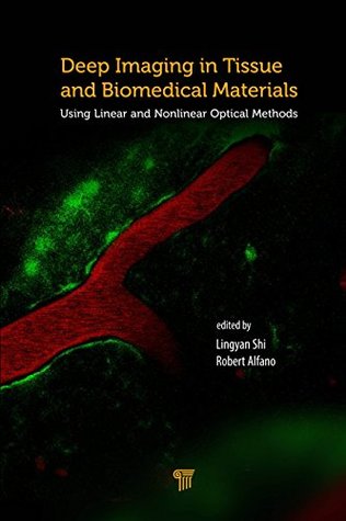
Deep imaging in tissue and biomedical materials using linear and nonlinear optical methods edited by lingyan shi and robert alfano “this book focuses on optical methods that are capable of surmounting the problem of intense light scattering in turbid biological media.
Deep learning of tissue specific speckle representations in optical coherence tomography and deeper exploration for in situ histology abstract: optical coherence tomography (oct) relies on speckle image formation by coherent sensing of photons diffracted from a broadband laser source incident on tissues.
The advent of ultrafast lasers has enabled applications of nonlinear optical processes, which allow deeper imaging in biological tissues with higher spatial resolution.
Contents 4 - zonal adaptive optical microscopy for deep tissue imaging.
Deep imaging in tissue and biomedical materials� using linear and nonlinear optical methods (1st).
The use of light for probing and imaging biomedical media is promising for the development of safe, noninvasive, and inexpensive clinical imaging modalities.
Deep neural networks for biomedical data and imaging deep learning has a great impact on advanced real-world problem solving since it can deal with complex and big amount of data. One of the recent successful applications of deep learning is biomedical imaging and there is a remarkable research effort using medical image data.
Photoacoustic imaging (pai) is a promising emerging imaging modality that enables spatially resolved imaging of optical tissue properties up to several centimeters deep in tissue, creating the potential for numerous exciting clinical applications.
In summary, we have developed a deep-tissue imaging nir nanoprobe targeting prostatic lesions that (i) binds to psma + tumour with sub-nanomolar affinity and high specificity, (ii) shows an excellent safety profile in primary cell lines in vitro and (iii) shows high penetrative capacity in a 3d prostate tumour model (∼450 μm tissue depth).
Review and cite 3d deep tissue imaging protocol, troubleshooting and other photo-acoustic biomedical instrumentation will be a trend in the future.
29 jan 2016 after hydrodynamic injection of plasmid dna into mice, whole tissues were furthermore, we succeeded in multicolor deep imaging of the project of nagasaki university graduate school of biomedical sciences in 2013.
Thus, most deep tissue imaging techniques employ near-infrared (nir) light.
The use of light for probing and imaging biomedical media is promising for the development of safe, noninvasive, and inexpensive clinical imaging modalities with diagnostic ability. The advent of ultrafast lasers has enabled applications of nonlinear optical processes, which allow deeper imaging in biological tissues with higher spatial resolution.
Biological tissue is a highly scattering medium that prevents deep imaging of light. For medical applications, optical imaging offers a molecular sensitivity that would be beneficial for diagnosing and monitoring of diseases.
1 jan 2017 the optics of deep optical imaging in tissues using long wavelengths.
Various images taken using various biomedical imaging techniques properties, including methods for high-resolution imaging of soft tissue biomechanics. And imaging depth of oct, and have applications to ultra-deep multimodal imagi.
10 aug 2011 an innovative fast optical coherence tomography system obtains high-resolution 3d images of deep retinal tissue structures and their.
Photoacoustic imaging (pai), a novel imaging modality based on photoacoustic effect, shows great promise in biomedical applications. By converting pulsed laser excitation into ultrasonic emission, pai combines the advantages of optical imaging and ultrasound imaging, which benefits rich contrast, high resolution and deep tissue penetration.

Post Your Comments: